So I found another SWEET blog The Weird Veterinary World (http://www.drdolen.com/). Dr Dolen has some amazing cases posted in there as well as very cool pictures!
Also, I had my first midterm on Monday in Clin Med I. It went very well, lots of basic husbandry questions as well as terms and definitions. Stuf like:
"What is a female cattle called that has not yet had a calf?"
Answer: Heifer
"What is the near-side of the horse?"
Answer: Left side
"What lympnh node can be palpated on HEALTHY animals?"
Answer: Submandibular
"What knot do you use when you need to lengthen a lead?"
Answer: Reef knot
"What part of the 'hay' contains the most fibre?"
Answer: Stem
Blah, blah, blah... Everyone feels like they over studied. Hopefully marks come up soon.
Histology quiz (worth 10%) is coming up soon... next Monday in fact. And just my luck, this weekend is my trip to Cornell University in Ithica, New York for the Veterinary Fraternity Grand Council 2011. :( Which means I have until Friday to study because I intend on being very drunk this weekend! Especially because I am legal to drink in the States! And also free keg beer!!
So what is histo.... well, the study of cells and tissues basically. It is cell bio on drugs... lots of purple, pink, blue, and black stained slides of every cell type from every organ... and the differences between species! Fun times. This test will be stains, epithelium, glands, muscles, nerves, and connective tissue.
 |
| Portion of the small intestine's villi. Note the simple columnar epithelial cells. The blue (basophilic) ovals are the parallel and polar nuclei of each columnar cells. The bright pink (eosinophilic) outline is called the brush/striated border and consists of microvilli. The small clear spots are mucous producing goblet cells. |
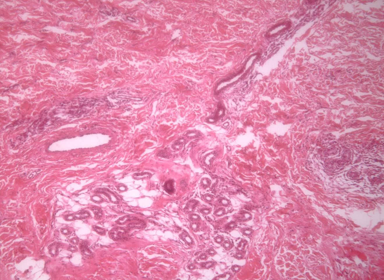 |
| This is a picture of a simple coiled tubular gland. It's actually an apocrine sweat gland of a dog. Simple cuboidal epithelial cells surround the gland's lumen. |
 |
| Striated muscle cells. You can see the dark lines and light lines interchange. Between the dark or Z lines is a sarcomere. A sarcomere is the contractile unit in the muscle. It contracts because of a Calcium dependant reaction between actin and myosin filaments. |
There we are. Hopefully I will have pictures of the Cornell Veterinary School's campus to post for you later. If I don't lose too many neurons this weekend!!!
No comments:
Post a Comment